Step 1: Receptor
Reading a protein structure, choosing which chains to keep, protonating and charging it, and deciding what to strip out before docking.
Docking scores a protein against a small molecule, and the score is only as good as the protein you hand it. A structure straight from a crystallographic database is not ready: it has no hydrogens, it may carry ordered waters and buffer molecules that were never part of the biology, it usually contains the ligand that was crystallised with it, and it has no partial charges at all. Step 1 turns that raw structure into a receptor.
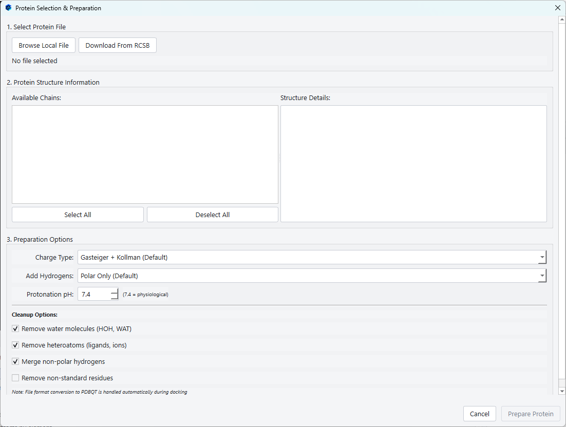
Load Receptor opens the Protein Selection and Preparation dialog, which does the whole job in one place: it reads the file, reports what is in it, takes your decisions about what to keep, and then runs the preparation before it closes.
1. Selecting a protein file
Two routes into the dialog, and they end in the same place.
Browse Local File
Opens a file chooser for a structure you already have. The receptor is loaded from a PDB file, so save a structure held in another format as PDB first. If you have been sent a model from a colleague, a homology model, or a structure you have already cleaned up in another program, this is the way in.
Download From RCSB
Asks for a four character Protein Data Bank identifier and fetches the structure directly, so a study can begin from nothing but an accession code.
2. Reading what the structure contains
As soon as a file is loaded, the middle of the dialog fills with two panels. On the left is the chain list, one entry per chain with its residue and atom count. On the right is a summary of the whole file.
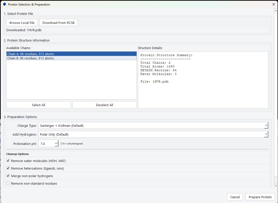
Read the summary before you go any further, because it tells you what decisions you are about to make.
| Reported figure | Why it matters |
|---|---|
| Total Chains | A structure with several chains may be a genuine multimer, or it may be several copies of the same molecule in the crystal. Docking into all of them at once is almost never what you want: pick the chain that carries the site you care about. |
| Total Atoms | A rough size check. If this is far smaller than you expected, the file may be a fragment or a single domain rather than the whole protein. |
| HETATM Records | Everything that is not a standard residue: the co-crystallised ligand, cofactors, metals, and buffer components. Some of these belong in your receptor and some do not. |
| Water Molecules | Ordered waters. Most are irrelevant to docking, but a few in the binding site can mediate a genuine hydrogen bond between the protein and the ligand. |
Choosing chains
Every chain is selected by default. Click to select just one, hold Ctrl to add or remove individual chains, and use Select All and Deselect All for the ends of the range. Only the chains you leave selected reach the prepared receptor, so deselecting the ones you do not need makes both the preparation and the docking faster.
3. Preparation options
Charge Type
The scoring function needs a partial charge on every atom. Four choices, and the default suits almost every case.
| Option | What it does | When to choose it |
|---|---|---|
| Gasteiger | Empirical charges computed from connectivity and electronegativity. | A general purpose scheme that works on anything, including non standard residues. |
| Kollman (United Atom) | Force field charges taken from a standard residue library. | Proteins made only of standard amino acids, where the library values apply directly. |
| Gasteiger + Kollman (Default) | Library charges where they exist, computed charges everywhere else. | The default, and the right answer for nearly every structure: standard residues get the well tested values and anything unusual is still charged. |
| None (Keep Original) | Assigns nothing and keeps whatever the file carried. | Only when the file already has charges you deliberately want preserved. |
Add Hydrogens
Crystal structures rarely contain hydrogens, but hydrogen bonding is most of what a docking score measures, so they have to be added.
| Option | Meaning |
|---|---|
| All Hydrogens | Adds every missing hydrogen, polar and non polar alike. |
| Polar Only (Default) | Adds only hydrogens on oxygen, nitrogen and sulfur, which are the ones that can donate a hydrogen bond. Non polar hydrogens are folded into the heavy atom they sit on. This is the standard preparation for docking and is why it is the default. |
| None (No Hydrogens) | Adds nothing. Only appropriate when the structure already has a complete and deliberate hydrogen set. |
Protonation pH
Sets the pH at which ionisable groups are protonated. The default of 7.4 is physiological and is what you want unless you are studying a compartment that is genuinely more acidic, such as a lysosome or a tumour microenvironment, in which case lower it accordingly.
4. Cleanup options
| Option | Default | What it removes and why |
|---|---|---|
| Remove water molecules (HOH, WAT) | On | Strips every ordered water. Most crystallographic waters are not part of the binding event, and leaving them in blocks parts of the site the ligand could otherwise occupy. Turn it off only when you have reason to believe a specific water mediates the interaction. |
| Remove heteroatoms (ligands, ions) | On | Strips the co-crystallised ligand, buffer molecules and ions. The ligand in particular must go, because it is sitting in exactly the site you are about to dock into. |
| Merge non-polar hydrogens | On | Folds hydrogens on carbon into the carbon itself. This is the united atom convention the scoring function was parameterised with, so leaving it on is correct. |
| Remove non-standard residues | Off | Strips modified residues such as phosphorylated serine or selenomethionine. Leave it off unless a modified residue is genuinely an artefact, because a modified residue in the site is usually part of the biology you are studying. |
Keeping waters in the binding site
Turning Remove water molecules off reveals a block of options for hydrated docking, which is the practice of keeping the few waters that genuinely bridge the protein and the ligand while discarding the rest.
| Control | What it does |
|---|---|
| Keep waters | Either distance based, which keeps only waters near the site, or all waters, which keeps every one in the selected chains. |
| Distance | The threshold for the distance based mode, from 1 to 15 Å. The default of 5.0 Å matches standard hydrated docking practice. |
| Binding site | Names the co-crystallised ligand that defines where the site is. Waters within the threshold of that ligand are kept and the rest are discarded. |
5. Preparing
Prepare Protein becomes available once a file is loaded and at least one chain is selected. Pressing it runs the preparation inside the dialog, with a progress bar, a stage label naming what is happening, and a log tail from the underlying tools.
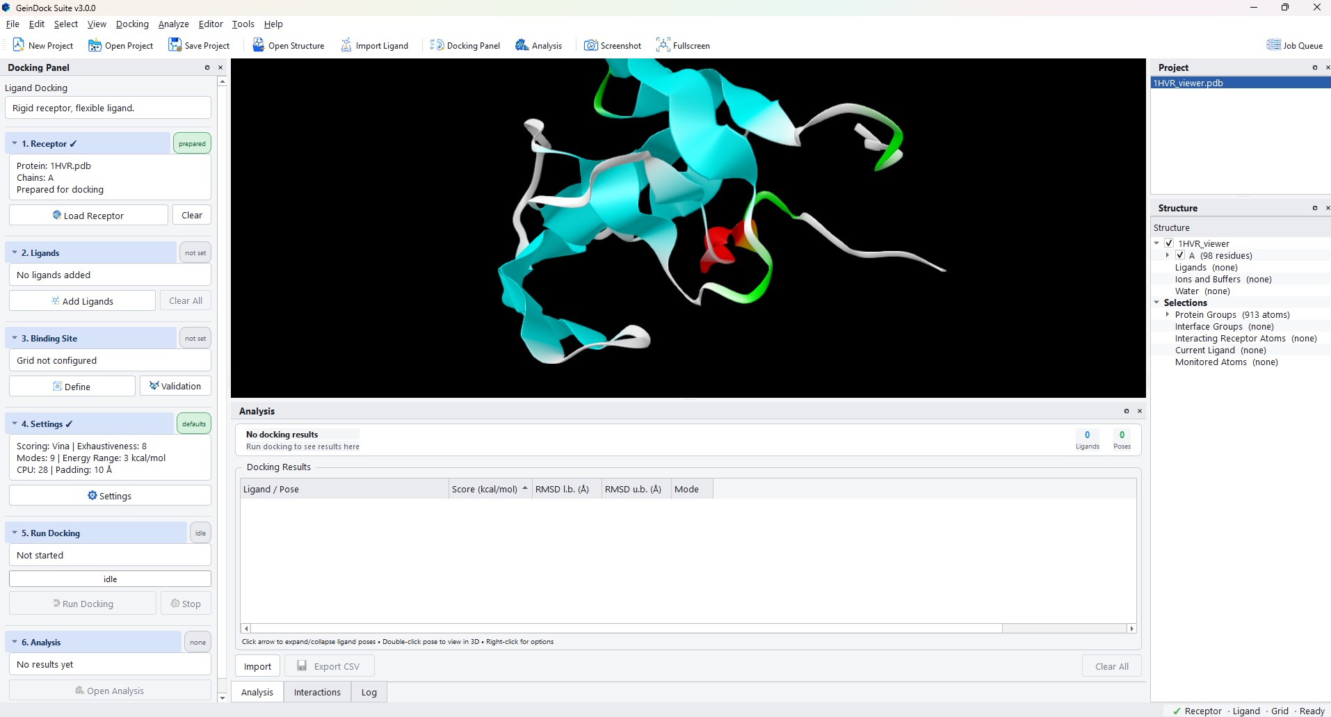
The preparation writes several files into the project workspace: a chain filtered copy for the viewer, a cleaned structure for building complexes after docking, and the charged, protonated receptor the docking search reads. You never need to open any of them; the summary in step 1 simply reports Prepared for docking when they exist.
Replacing or clearing the receptor
Clear forgets the current receptor and its prepared files, returning step 1 to not set. Loading a different receptor over the top has the same effect. Either way the binding site from the old receptor no longer describes anything meaningful, so redefine it in step 3 before you run again.