Ligand docking
The default protocol: a rigid receptor and a flexible ligand. What the search explores, what it holds fixed, and how to read what comes back.
Ligand docking is the default protocol and the one every other mode is built on. The receptor is held rigid, the ligand is free to rotate about its rotatable bonds, and the search looks for the placement inside the box that the scoring function likes best.
The panel heading reads Ligand Docking with the note Rigid receptor, flexible ligand, and step 4 is Settings.
What the search actually explores
Three things vary during the search, and it is worth being clear about which they are, because everything the protocol can and cannot do follows from the list.
| Varies | Held fixed |
|---|---|
| Where the ligand sits inside the box, in all three directions. | Every receptor atom. Nothing on the protein moves. |
| How the ligand is oriented. | The ligand's bond lengths and bond angles. |
| The torsion angle of each of the ligand's rotatable bonds. | The ligand's ring conformations and its stereochemistry. |
Running it
The whole procedure is the six steps of the Docking Panel, in order. There is nothing mode specific to do.
- Prepare the receptor. Step 1. Choose the chain that carries your site, strip the waters and heteroatoms, and let it protonate at pH 7.4.
- Prepare the ligands. Step 2. Check the atom counts and the validity column before you move on.
- Place the box. Step 3. Use the crystal ligand if the structure had one, and read the clearance note before you commit.
- Prove the box. Validation. Once per target, this tells you whether the configuration can reproduce a pose that is known to be right.
- Choose a precision. Step 4. Swift for a first look or a screen, Rigorous for anything you intend to report.
- Run it and read the poses. Step 5 and the pose table. Sort by score, then look at whether the top poses agree with one another.
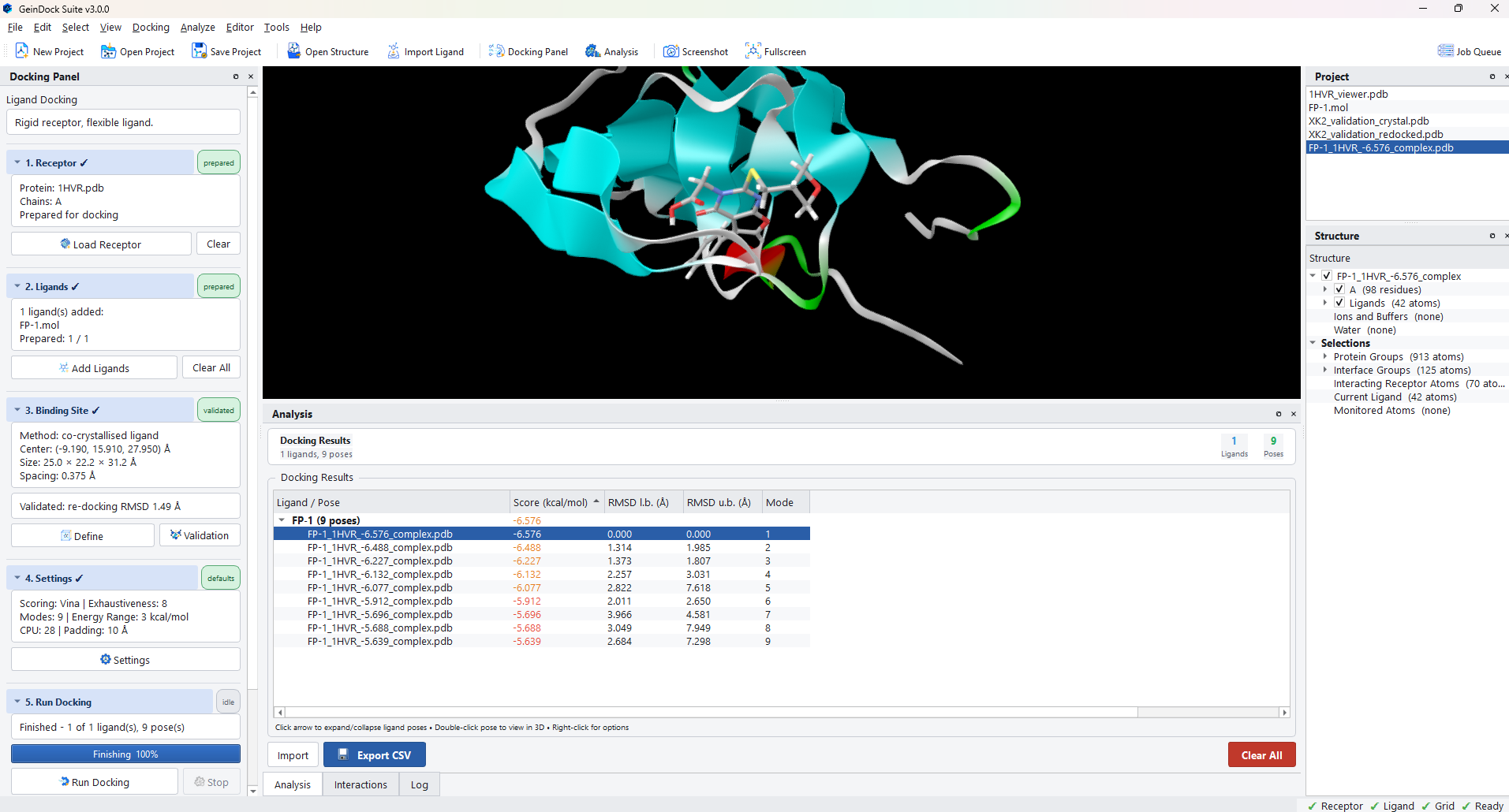
What the columns mean for this mode
The pose table carries columns for every mode, and the ones that do not apply stay empty. In ligand docking the ones that matter are these.
| Column | What it tells you |
|---|---|
| Score (kcal/mol) | The scoring function's estimate of binding affinity. More negative is better. It is an estimate from an empirical function, not a measurement, so use it to rank compounds against each other rather than to predict a number. |
| RMSD l.b. (Å) | How far this pose is from the best pose, measured conservatively. The best pose is 0 by definition. |
| RMSD u.b. (Å) | The same comparison measured strictly. A large gap between the two bounds means the pose is similar in shape but differently oriented, which is often a symmetric molecule flipped end for end. |
| Mode | The rank of the pose within its ligand, 1 being the best scoring. |
When ligand docking is the wrong tool
The rigid receptor is the assumption that limits this mode, and there are three situations where it breaks down.
- The pocket rearranges on binding. A side chain that has to swing aside to let a compound in will instead reject it, because in a rigid receptor it is a wall. Use flexible docking.
- You know something the search keeps missing. If a series is known to hinge on a particular contact and the search will not make it, stating the requirement is more honest and more useful than raising exhaustiveness until it happens by chance. Use constraint docking.
- The compound reacts with the protein. A covalent inhibitor scored as though it were sitting freely in the pocket is being scored for the wrong thing entirely. Use covalent docking.