Box, solvent, ions and membranes
The periodic box, the water model, the salt and, for membrane proteins, the lipid bilayer and its staged equilibration.
The box
The complex is placed in a periodic box so that it is surrounded by solvent on every side. The shape and the solute-box distance decide how much water the system holds, and with it the speed of the run.
| Shape | Characteristic | When to use it |
|---|---|---|
| "Rhombic dodecahedron" (default) | The closest shape to a sphere among periodic boxes; needs the least water for a given distance. | Almost always: a globular complex tumbling freely. |
| "Truncated octahedron" | Also close to a sphere, slightly more water than the dodecahedron. | When a later tool expects this shape. |
| "Cubic" | Simple to picture, but the most water for the same distance. | When a simple box is required, or for a comparison with older work. |
| "Rectangular (triclinic)" | Builder only: a box with the edges of the solute plus the distance. | An elongated solute whose orientation is kept by restraints. |
"Solute-box distance" (default 1.0 nm; 0.8 to 2.0 nm on Run MD, 0.5 to 5.0 nm on the Builder) is the smallest distance between the solute and the box edge. With the 1.0 nm cut-off, 1.0 nm keeps a solute from interacting with its own periodic image through the cut-off as long as it does not change shape much. Increase it for a flexible protein that may unfold or extend, or for a complex with long termini.
Water
Run MD and the Batch queue use TIP3P water, the model AMBER ff14SB and GAFF2 were parametrised with. The System Builder lists the water models of the chosen force field; with amber14sb these are OPC, OPC3, SPC, SPC/E, TIP3P and TIP4P-Ew. Change the model only for a deliberate comparison: the protein and ligand parameters are balanced against TIP3P.
Ions
| Setting | Default | Meaning and when to change it |
|---|---|---|
| "Neutralise" | on | Counter-ions bring the net charge of the system to zero, which PME electrostatics assumes. Leave it on. |
| "Salt" / "Salt concentration" | 0.15 mol/L NaCl (0 to 2) | Added after neutralisation; 0.15 mol/L is physiological. Set 0 for a comparison with work in pure water, or the buffer concentration of an experiment you want to match. |
| "Ions" (Builder) | NA and CL | The ion names of the force field; SOD and CLA with CHARMM36. |
Ions replace randomly chosen water molecules at least 0.6 nm from the solute. The random choice uses the seed 1993, so the same input always gives the same system.
Membrane proteins
A membrane protein in a box of water is not in its native environment: its hydrophobic surface faces water, and loops and helices can move in ways the bilayer would not allow. Geinium therefore checks every protein before the start. A protein with two or more segments whose mean Kyte-Doolittle hydropathy over a 19-residue window is above 1.6 is treated as a membrane protein. If no membrane was chosen, the "Membrane protein" dialog asks for a decision.

| Choice | What happens | When to use it |
|---|---|---|
| "Use a membrane (POPC:CHOL 7:3)" | The job is switched to a membrane system: the protein is embedded in a Lipid21 bilayer and the staged equilibration below is used. | Whenever the transmembrane part matters for the question. |
| "Simulate without a membrane" with "Reason (required)" | The protein is simulated in water. The reason is stored in the job and printed in the "Membrane" row of the report. | A deliberate choice, such as a study of an extracellular vestibule or a soluble domain, or a fast screen whose limits you accept. |
The dialog is asked by Run MD and the Batch queue before the start, and by the System Builder before a build. A report for a membrane protein simulated in water without a stated reason is blocked by the scientific checks of the export.
The bilayer
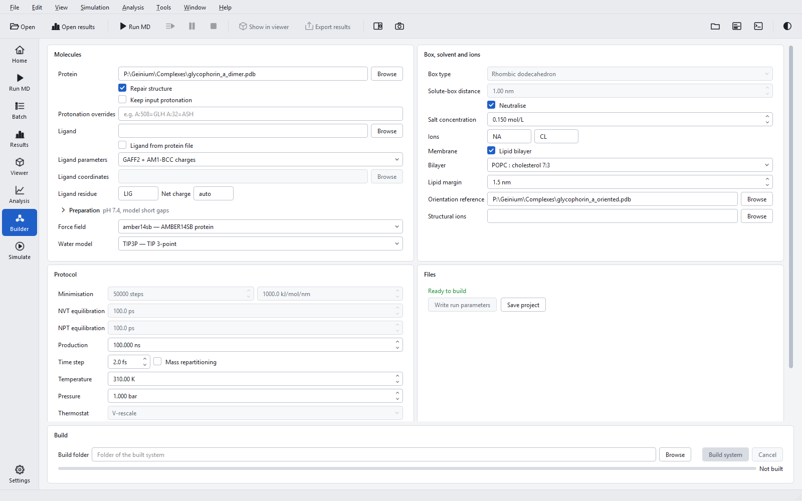
On Run MD and in the Batch defaults the check box "Membrane (POPC:CHOL 7:3)" embeds the protein in a bilayer of POPC and cholesterol in the ratio 7:3, built from the Lipid21 parameters. The System Builder offers the same with more control:
| Builder control | Default | Meaning and when to change it |
|---|---|---|
| "Lipid bilayer" | off | Embeds the protein in a bilayer. Needs AMBER ff14SB and TIP3P, and switches the temperature from 300 to 310 K. |
| "Bilayer" | "POPC : cholesterol 7:3" | Or "POPC": a pure POPC bilayer, simpler and faster to equilibrate, for test systems and for proteins studied in pure phosphatidylcholine. |
| "Lipid margin" | 1.5 nm | Lateral distance from the protein to the box edge, filled with lipids. Increase it for a protein whose surrounding lipids you want to analyse. |
| "Orientation reference" | empty | A structure of the same protein already oriented in a membrane (membrane normal along z), for example from a membrane-orientation reference database. Strongly recommended; see below. |
| "Structural ions" | empty | Ion positions in the frame of the orientation reference, for ions that belong to the protein structure. They are transferred and restrained. Requires the reference. |
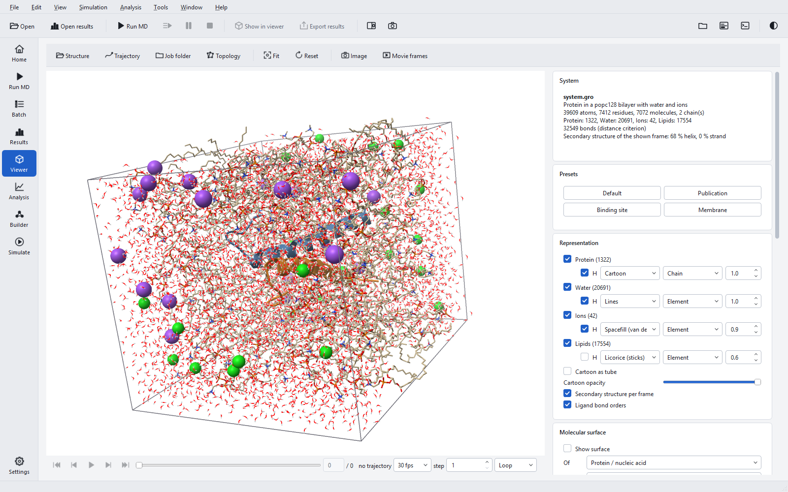
The bilayer is cut from pre-built Lipid21 patches. The POPC:cholesterol patch holds 70 POPC and 30 cholesterol per leaflet (7.286 x 7.286 x 8.526 nm, 47,232 atoms); the POPC patch holds 64 POPC per leaflet (32,540 atoms).
Orientation in the membrane
With an orientation reference, the C-alpha atoms of the protein are aligned to those of the reference and superposed, so the protein sits in the bilayer as the reference says. Without one, Geinium fits the hydrophobic belt of the protein to a slab and warns. The fit was within 3.5 degrees and 0.12 nm for a 544-residue transporter, but 20.6 degrees off for a small helix dimer, whose hydrophobic belt is too short to define the tilt well. Use a reference whenever one exists.
Construction checks
The protein keeps a lateral margin of 1.5 nm of lipids, at least 1.5 nm of water beyond the solute and 2.0 nm beyond the head-group planes. Lipids that overlap the protein are removed, the leaflets are balanced to within 5 %, and no water is left in the hydrophobic core. Before the system is accepted, the closest heavy-atom distance, lipid rings pierced by other molecules, the net charge and the leaflet balance are checked; a system that fails is refused rather than simulated.
The membrane protocol
A protein freshly embedded in a bilayer has voids around it and lipids that do not yet pack against it. A membrane system therefore replaces the NVT and NPT equilibration of a soluble system with six stages that release restraints step by step. Minimisation runs for 5000 steps. All stages use 310 K, a thermostat time constant of 1.0 ps, and from the third stage on semi-isotropic C-rescale pressure coupling with a time constant of 5.0 ps, so the area and the thickness of the bilayer relax independently.
| Stage | Ensemble and length | Time step | Backbone / side-chain restraints (kJ/mol/nm2) | Lipid head-group height | Lipid dihedrals (kJ/mol/rad2) |
|---|---|---|---|---|---|
| Equilibration 1 | NVT, 125 ps | 1 fs | 4000 / 2000 | 1000 | 1000 |
| Equilibration 2 | NVT, 125 ps | 1 fs | 2000 / 1000 | 400 | 400 |
| Equilibration 3 | NPT, 250 ps | 1 fs | 1000 / 500 | 400 | 200 |
| Equilibration 4 | NPT, 1 ns | 2 fs | 500 / 200 | 200 | 200 |
| Equilibration 5 | NPT, 2 ns | 2 fs | 200 / 50 | 0 | 100 |
| Equilibration 6 | NPT, 5 ns | 2 fs | 50 / 0 | 0 | 0 |
Together these are 8.5 ns per replica. The equilibration lengths, minimisation settings, thermostat and barostat of a soluble job do not apply to a membrane job. With 4 fs chosen, hydrogen-mass repartitioning is used in production only and equilibration keeps its 1 and 2 fs; 1 fs production is not accepted for membranes.
Rebuilding a system into a folder that already holds simulation output moves the old output aside to <name>_before_rebuild_<time> instead of overwriting it.